Existing Patients
(740) 344-4549
New Patients
(740) 212-1897

At the office of Brian Howe DDS, Family Dentistry, we leverage cone-beam computed tomography (CBCT) to gather clear, three-dimensional information about the teeth, jaws, and surrounding structures. CBCT is a focused imaging tool designed for dental and maxillofacial needs — it reveals anatomical detail that standard two-dimensional X-rays cannot, helping clinicians see relationships and subtleties that influence accurate diagnosis and treatment planning.
Our approach is to use CBCT selectively and purposefully, applying it when the additional information will materially improve care. The images produced by modern CBCT units are fast to acquire and provide clinicians with practical views that support more predictable outcomes across a wide range of dental procedures.
Cone-beam CT creates volumetric scans that let clinicians examine teeth, bone, nerves, and sinus anatomy from multiple angles. Rather than a flattened representation, these scans present depth and spatial relationships — for example, the exact position of a root relative to the mandibular nerve or the thickness of bone in a potential implant site. That extra perspective reduces guesswork when planning care.
In orthodontics and surgical planning, CBCT helps reveal tooth impactions, eruption patterns, and the orientation of roots that may be obscured on standard films. For patients with unusual anatomy or complex restorative needs, a 3D dataset can clarify whether a minimally invasive approach is possible or if more involved treatment is required.
CBCT also improves the ability to screen for bony pathology and developmental anomalies. While radiographs remain useful for routine exams, the selective use of CBCT is an important diagnostic step when a detailed assessment of the hard tissues will affect the clinical decision-making process.
Detailed CBCT images allow clinicians to plan procedures with measurable precision. For example, when assessing bone volume and angulation for implant placement, a three-dimensional view lets the team choose optimal implant size, position, and orientation to support long-term function and esthetics. This level of planning reduces surprises during treatment.
Endodontic diagnosis is another area where CBCT can be invaluable. Scans can reveal additional canals, root fractures, or complex anatomy that may be missed on periapical films. Identifying these issues before treatment helps tailor techniques to the case at hand and can improve the chances of successful outcomes.
Beyond surgical planning, CBCT datasets support restorative work by clarifying occlusal relationships and prosthetic space. When combined with clinical examination and digital impressions, the information from a CBCT scan contributes to a comprehensive, predictable treatment plan.
Implant treatment benefits directly from CBCT because the scans allow surgeons to virtually place implants and evaluate surrounding anatomy before entering the surgical site. Viewing the proximity to nerves, sinus cavities, and adjacent teeth helps determine whether bone grafting is necessary and where to position implants for reliable support and esthetic harmony.
For impacted or surgically complex extractions, cone-beam imaging brings clarity about root morphology and the relationship of roots to vital structures. That clarity supports surgical strategies that minimize trauma, preserve bone, and promote faster healing after the procedure.
When oral surgery is coordinated with restorative goals, CBCT provides a shared reference for both the surgical and restorative teams. This collaborative planning improves communication and aligns surgical steps with the desired prosthetic outcome.
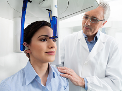
CBCT scans are quick — typically completed in less than a minute of acquisition time — and the process is noninvasive. Patients remain seated or standing while the scanner rotates around the head, creating a volumetric image with minimal movement required. The short scan time contributes to a comfortable experience for most patients.
Modern dental CBCT units are designed with focused fields of view and protocols that limit radiation exposure to what is necessary for the diagnostic task. Clinicians follow the principle of ALARA (as low as reasonably achievable) when deciding to image, ensuring that each scan is justified by a specific clinical need.
Before any CBCT scan, our team reviews the indications and explains what the scan will show and how it will be used in planning care. This conversation helps patients understand the value of the imaging and what to expect during and after the appointment.
Acquiring a CBCT scan is only one part of the process; interpreting the three-dimensional dataset correctly is essential. Our clinicians review scans in the context of the patient’s history, clinical exam findings, and treatment goals. When complex or unusual findings appear, we consult with specialists or imaging experts to ensure a comprehensive interpretation.
Because CBCT reveals anatomical detail that may not be visible clinically, follow-up steps can include targeted clinical checks, additional imaging, or referrals when appropriate. The goal is always to translate imaging information into a clear, actionable plan that respects the patient’s needs and preferences.
Ultimately, CBCT is a tool that improves diagnostic clarity and helps clinicians deliver care with greater confidence. If you have questions about how three-dimensional imaging might apply to your dental care, please contact us for more information. Our team will gladly explain the role of CBCT in your treatment and help determine whether it is the right choice for your situation.
Cone-beam computed tomography, commonly called CBCT, is a three-dimensional imaging technology used in dentistry to visualize teeth, jawbone, nerves and sinuses with high spatial detail. A rotating scanner captures a volumetric dataset in a single pass, and specialized software reconstructs that data into cross-sectional and 3D views clinicians can examine from multiple angles. Because CBCT acquires volumetric information rather than a flat image, it reveals spatial relationships and depth that conventional two-dimensional radiographs cannot.
Clinicians use CBCT datasets to measure bone dimensions, evaluate root anatomy, and assess anatomical variations that influence diagnosis and treatment planning. The images can be reformatted into slices, panoramic views, and 3D renderings to communicate findings and options clearly. Interpretation combines the scan with the clinical exam to ensure imaging findings are correlated with symptoms and treatment goals.
CBCT is recommended when a three-dimensional assessment will materially affect diagnosis or treatment decisions, such as implant planning, evaluation of impacted teeth, assessment of complex root anatomy, or investigation of suspected bony pathology. It is used selectively rather than routinely, applied when the extra information improves safety, predictability, or overall outcomes. Your clinician will weigh the expected diagnostic benefit against the need to limit radiation exposure.
Common indications include preoperative implant evaluation, assessment of failed endodontic therapy, analysis of facial trauma or fractures, and detailed surgical planning for extractions or orthognathic procedures. In orthodontics, CBCT can clarify eruption patterns and skeletal relationships in complex cases. The decision to image is guided by clinical findings, patient history, and the specific questions the team needs answered.
Traditional dental X-rays, such as bitewings and periapical films, produce two-dimensional images that are excellent for routine screening of cavities and periodontal bone levels. CBCT, by contrast, produces a volumetric dataset that preserves depth, allowing clinicians to view structures in three dimensions and to slice through anatomy in any plane. That difference is particularly important when assessing relationships to nerves, sinus cavities, or the true volume of bone for implant placement.
While two-dimensional radiographs remain useful for many everyday diagnostic tasks, CBCT offers complementary information when anatomy is complex or when precise spatial data is required. The technologies are used together: conventional films for routine monitoring and CBCT for targeted diagnostic challenges. Choosing the right imaging modality depends on the clinical question and the level of detail needed.
A CBCT scan is quick and noninvasive. Most modern units complete the image acquisition in less than a minute while the patient sits or stands still and the scanner rotates around the head. The staff will position you carefully, explain the process, and instruct you to remain still for the brief scan to minimize motion artifact.
Preparation is minimal; you may be asked to remove glasses, jewelry or removable dental appliances that could interfere with imaging. After acquisition, the dataset is reviewed and processed by the clinical team, who will explain what the images show and how they will inform the treatment plan. If additional consultation with a specialist is warranted, the team will coordinate next steps.
CBCT uses ionizing radiation, so safety protocols are applied to minimize exposure while obtaining diagnostically useful images. Modern dental CBCT units offer focused fields of view and adjustable imaging protocols so clinicians can limit the scanned area to what is necessary for the clinical task. The principle of ALARA (as low as reasonably achievable) guides imaging decisions to balance diagnostic benefit with radiation stewardship.
Your dental team will select the smallest field of view and lowest acceptable dose that still answers the clinical question, and they will avoid unnecessary repeat scans. For vulnerable populations, such as children or pregnant patients, extra precautions are taken and alternative imaging strategies are considered when appropriate. If you have concerns about radiation, discuss them with your clinician so they can explain the specific benefits and safeguards in your case.
CBCT provides precise measurements of bone height, width and angulation, enabling clinicians to virtually plan implant position, size and orientation before surgery. The ability to visualize the exact relationship of proposed implant sites to nerves, adjacent teeth and sinus cavities reduces intraoperative surprises and helps determine whether bone grafting or staged procedures are necessary. This 3D information supports surgical guides and more predictable implant placement.
By integrating CBCT data with clinical findings and digital impressions, the restorative and surgical teams can coordinate toward an outcome that balances function and esthetics. Virtual planning can also simulate prosthetic space and occlusal relationships, helping to ensure implant position supports the desired final restoration. This collaborative approach often results in fewer complications and a more streamlined treatment sequence.
Yes. Because CBCT visualizes bone and surrounding anatomy in three dimensions, it can reveal cysts, tumors, developmental anomalies, root fractures, and areas of bone destruction that may be obscured or hidden on two-dimensional films. Small lesions or complex anatomical variations can be more readily identified and localized with a volumetric scan. Detecting these issues early can influence treatment decisions and prompt timely referral when appropriate.
CBCT is particularly useful when conventional imaging and the clinical exam do not fully explain symptoms such as unexplained pain, swelling, or persistent infection. Interpretation always considers the entire clinical picture; incidental findings that require monitoring or referral are documented and discussed with the patient. When necessary, the team will collaborate with oral and maxillofacial radiologists or other specialists for a more detailed review.
CBCT scans are reviewed by your treating dentist in the context of your medical and dental history, the clinical examination, and the treatment objectives. When scans show complex or unusual findings, the office may consult with or refer images to an oral and maxillofacial radiologist or a specialist who has advanced training in interpreting three-dimensional imaging. This collaborative approach helps ensure a comprehensive and accurate diagnosis.
After interpretation, the clinician will explain the findings, outline recommended next steps, and describe how the imaging information affects proposed treatments. Results can lead to targeted clinical checks, adjustments in surgical or restorative plans, or referrals to specialists. Clear communication ensures you understand how the scan informs your care and what to expect moving forward.
CBCT is not always required for routine dental care; simple cavities, routine periodontal screenings and basic restorative assessments are typically managed with conventional radiographs and a clinical exam. Imaging is reserved for situations where three-dimensional information will change management or improve safety. Using CBCT indiscriminately is avoided to reduce unnecessary radiation exposure and to keep care focused and efficient.
Certain patient conditions, such as inability to remain still during the scan or contraindications related to pregnancy, may prompt clinicians to defer CBCT or select alternative approaches. When the need for CBCT is uncertain, the team will discuss other diagnostic options and the potential diagnostic yield so patients can make informed decisions about their care.
At the office of Brian Howe DDS, Family Dentistry, CBCT is used selectively to enhance diagnostic clarity and support precise treatment planning across implant dentistry, endodontics, oral surgery and complex restorative cases. The team integrates volumetric imaging with clinical examination and digital workflows to tailor care to each patient’s anatomy and goals. This measured use of technology helps reduce surprises during procedures and contributes to more predictable results.
When CBCT is indicated, clinicians explain the reason for imaging, review the results with the patient, and incorporate findings into a coordinated treatment plan that prioritizes safety and long-term function. For complex interpretations, the practice consults with imaging specialists to ensure comprehensive analysis. Patients are encouraged to ask questions so they understand how CBCT findings influence their personalized care pathway.
Our friendly and knowledgeable team is always ready to assist you. You can reach us by phone at (740) 344-4549 or by using the convenient contact form below. If you submit the form, a member of our staff will respond within 24–48 hours.
Please do not use this form for emergencies or for appointment-related matters.
