Existing Patients
(740) 344-4549
New Patients
(740) 212-1897

Digital impressions have changed how dentists capture the shape, position, and texture of teeth and adjacent soft tissues. Instead of traditional putty-based molds, intraoral optical scanners create a precise, three-dimensional digital model that clinicians can view, manipulate, and share instantly. For patients, that means a more comfortable exam experience; for the clinical team, it means clearer communication, fewer remakes, and streamlined workflows. At the office of Brian Howe DDS, Family Dentistry, this technology supports everything from routine crowns to advanced restorative care.
At their core, digital impressions are high-resolution scans of the mouth captured with intraoral cameras and optical sensors. These devices gather thousands of data points per second and stitch them together to produce an accurate 3D model of a patient’s dentition and surrounding tissues. The result is a virtual replica that can be used for diagnosis, treatment planning, restorative design, and laboratory fabrication — all without the discomfort or mess associated with conventional impression materials.
Because the dataset is digital, it preserves fine details such as margin lines, occlusal contacts, and interproximal contours that are critical for long-lasting restorations. Clinicians can zoom, rotate, and measure directly on-screen, making it easier to confirm fit and form before any restoration is fabricated. This level of precision reduces the likelihood of adjustments and remakes once a restoration arrives from the lab.
Beyond immediate clinical benefits, digital impressions integrate with other digital tools — including CAD/CAM design software, cone beam CT, and practice management systems — enabling a cohesive digital workflow. That interoperability creates efficiencies that benefit the entire care team and, ultimately, the patient’s overall experience and outcome.
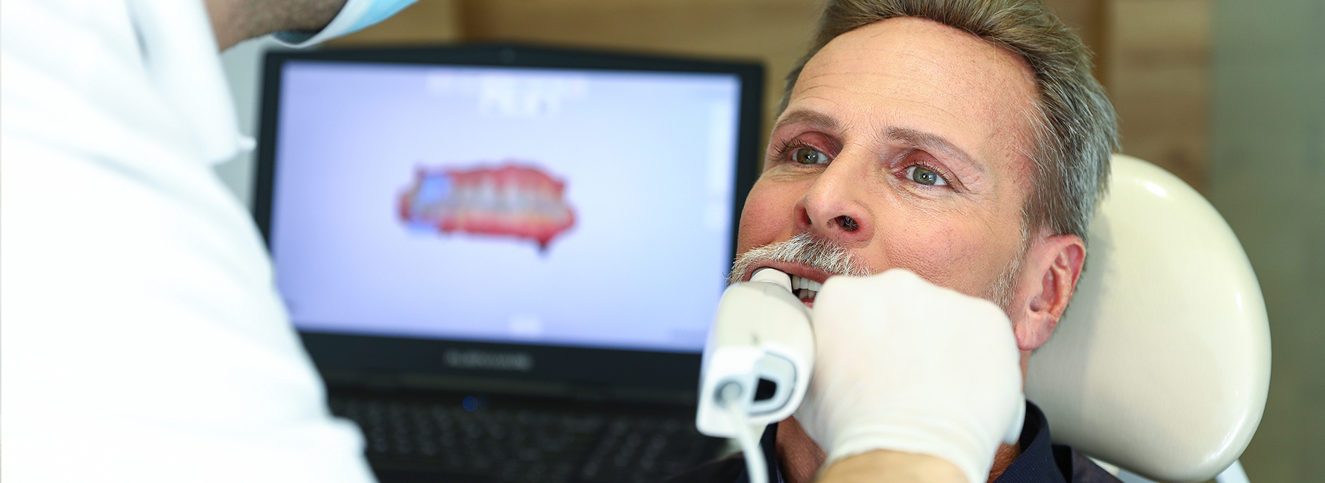
Intraoral scanning begins with a technician or dentist moving a wand-like scanner around the teeth and gums. The scanner emits light and captures reflected images, which proprietary software rapidly processes into a continuous, three-dimensional model. The process is noninvasive and typically takes only a few minutes for most cases, depending on the area being scanned and the complexity of the restoration.
During the scan, the clinician can monitor progress on a chairside display and identify any areas that need rescanning in real time. This immediate feedback is a major advantage over conventional impressions, where errors may not be visible until the material is removed and poured into a model. With digital scanning, common problems such as voids, distortions, or missed margins are easy to detect and correct on the spot.
Once the scan is complete, the digital file can be refined and annotated to highlight margin locations or special instructions for the dental laboratory. Files are exported in standardized formats compatible with most labs and manufacturing centers, which helps preserve accuracy throughout the restorative process.
Accuracy is a primary reason clinicians adopt digital impressions. High-resolution scans capture microscopic details that ensure crowns, bridges, and onlays seat properly and maintain healthy periodontal margins. Because the digital model reflects the true anatomy of the mouth, lab technicians can fabricate restorations that require fewer chairside adjustments, improving both longevity and patient satisfaction.
Digital impressions also support better diagnostic communication. With a shareable, manipulable 3D model, the dentist, lab technician, and patient can review the proposed outcome together. This transparency helps set realistic expectations and allows the clinician to make informed choices about materials, occlusal design, and esthetic considerations.
Predictability extends to implant planning and orthodontics as well. When combined with CBCT imaging or digital treatment planning software, digital impressions contribute to precise implant placement guides and aligner designs that track closely to the intended treatment path.
One of the most tangible benefits of digital impressions is reduced turnaround time. Instead of packaging and shipping physical impressions, clinicians transmit digital files electronically to dental laboratories or in-office milling units. That electronic handoff shortens lab cycles, minimizes the risk of damage or distortion in transit, and accelerates delivery of final restorations.
For practices equipped with chairside CAD/CAM systems, digital impressions are the foundation for same-day restorations. The digital scan feeds directly into design software, allowing the clinician to design a crown, mill it from a ceramic block, and deliver a final restoration within a single appointment. This capability can be a major convenience for patients who prefer fewer visits or have scheduling constraints.
Even when a lab-fabricated restoration is required, the streamlined communication and improved fit often translate into faster case completion and fewer follow-up visits. That smoother workflow improves practice efficiency and enhances the patient’s overall experience.
For many patients, comfort is the most immediate advantage of digital impressions. There’s no heavy tray filled with viscous material that can trigger gag reflexes or cause discomfort; instead, the scanning process feels similar to a routine intraoral camera examination. This patient-friendly approach makes it simpler to obtain accurate records from anxious or sensitive patients.
Digital files also change how clinicians communicate with patients and labs. Chairside visualizations allow patients to see and understand their treatment needs in three dimensions, improving informed consent and engagement. On the laboratory side, technicians receive clearer, more detailed data and can provide feedback or request refinements without waiting for physical models.
That collaborative dynamic supports better outcomes. Whether a case is routed to an outside lab or produced in-office, the shared digital model becomes a single source of truth — reducing misunderstandings and ensuring the final restoration aligns with the treatment goal.
Digital impressions are a modern tool that supports accurate restorations, quicker workflows, and a more comfortable patient experience. As part of a comprehensive digital strategy, they help clinicians make better decisions and deliver predictable results. If you’d like to learn more about how digital scanning is used in our practice or whether it’s right for your care, please contact us for more information.
Digital impressions are high-resolution, three-dimensional scans of the teeth and surrounding soft tissues captured with an intraoral scanner and specialized software. Instead of using putty-filled trays to make a physical mold, the scanner collects thousands of data points and stitches them into a precise virtual model. The resulting digital file can be reviewed, measured, and edited on-screen before being sent for restoration design.
Compared with traditional impressions, digital scans eliminate many common sources of distortion such as material shrinkage, voids, or tray movement. Clinicians can identify and correct errors immediately, which reduces the need for remakes and chairside adjustments. The digital format also makes it easier to integrate scans with CAD/CAM systems and laboratory workflows.
An intraoral scanner uses a small, wand-like tip that emits light and captures reflected images of the teeth and gums as the clinician moves it through the mouth. Proprietary imaging software processes those reflections in real time and assembles a continuous 3D model from overlapping frames. The scanner records fine surface detail including margins, contacts, and occlusal anatomy by collecting thousands of data points per second.
During the scan the clinician watches a chairside display to confirm full coverage and to identify any areas that need rescanning. If a detail is missed, the operator can recapture that region immediately so the final model is complete. The finished file is then refined, annotated if needed, and exported in standard formats for lab or in-office milling use.
Yes, modern digital impressions deliver the precision required for crowns, bridges, onlays, and implant restorations by capturing microscopic surface details and clear margin lines. High-fidelity scans allow technicians to design restorations that seat properly and respect periodontal contours, which helps maintain long-term function and tissue health. Multiple studies and clinical experience show that digitally produced restorations often require fewer adjustments at delivery.
When used alongside CAD/CAM manufacturing and quality laboratory protocols, digital scans support predictable restorative outcomes for both single-unit and multi-unit cases. For implant care, digital impressions combined with digital treatment planning create accurate surgical guides and final prosthetics. The clinician’s skill in capturing full-arch or multiple-segment scans remains an important factor in final accuracy.
Digital impressions are a key enabler of same-day restorations because the scan can be fed directly into chairside CAD/CAM design software. Once the design is finalized, an in-office milling unit can fabricate a ceramic crown or onlay during the same appointment, allowing the clinician to deliver the final restoration before the patient leaves. Eliminating shipping and model-pouring steps shortens turnaround time and reduces the number of visits for many cases.
Even when a case is routed to an external laboratory, the electronic transfer of a high-quality scan accelerates communication and reduces the risk of distortion during transit. The result is often faster lab cycles and fewer follow-up adjustments, which improves scheduling efficiency and enhances the overall patient experience. Cases requiring complex laboratory work or additional consultations may still need multiple visits, but the digital workflow streamlines each step.
During a digital impression appointment the clinician or trained team member will move a compact scanner around your teeth and gums while you sit comfortably in the chair. The procedure is noninvasive and typically takes only a few minutes for a single quadrant or a bit longer for full-arch captures. Many patients find the experience more pleasant than conventional impressions because there is no impression tray or putty and less likelihood of gagging.
At Brian Howe DDS, Family Dentistry clinicians review the scan on a chairside monitor and confirm that margins and occlusal relationships are captured clearly before concluding the appointment. If any area requires additional detail, the operator can rescan that spot immediately, avoiding delays later in the process. After capture, the digital file is refined and prepared for design, milling, or laboratory transmission according to the planned restoration.
Although digital impressions are suitable for most restorative and orthodontic cases, there are scenarios where conventional impressions or a hybrid approach may be preferred. Heavy bleeding, significant subgingival margins, extreme tissue retraction challenges, or very limited mouth opening can make intraoral scanning more difficult and may necessitate traditional techniques. Clinical judgment guides the choice of method to ensure the best outcome for complex conditions.
In some multi-step or highly customized laboratory workflows technicians may request a physical model or analog component, and clinicians will use the approach that yields the most predictable result. The goal is always to select the impression technique—digital, conventional, or both—that best supports accuracy and long-term success for the individual patient.
Digital impression files are exported in standardized formats such as STL, PLY, or other manufacturer-specific file types and then transmitted electronically to the chosen dental laboratory. File transfer typically occurs via secure, encrypted connections or through the lab’s protected portal to maintain fidelity and prevent corruption during transit. Clear file protocols and standardized formatting help labs work efficiently and preserve the detail clinicians captured.
Practices implement privacy and security protocols to protect patient data during storage and transmission, and many systems include user access controls and audit logs. Digital workflow partners and labs should follow industry best practices for data protection and comply with applicable privacy regulations. Patients with questions about how their records are handled can ask the practice team for details about security measures and consent procedures.
Digital impressions can be merged with cone beam CT (CBCT) scans and other digital records to create a comprehensive, three-dimensional view of both hard and soft tissues. This integration allows clinicians to plan implant placement, design surgical guides, and evaluate occlusion and esthetics within a single digital environment. The combined dataset improves coordination between restorative planning and surgical execution, increasing the predictability of outcomes.
For orthodontic and aligner therapy, accurate surface scans feed into treatment planning software that simulates tooth movement and produces aligner geometries. In implant workflows the merged data help generate precise implant guides and provisional restorations that reflect the planned prosthetic result. The interoperability of digital tools streamlines case planning and supports multidisciplinary collaboration.
Digital impressions create a single, shareable 3D model that serves as a common reference for dentists, laboratory technicians, and patients, which improves clarity during case planning. Chairside visualization lets clinicians show patients the scanned anatomy and proposed restorative areas, helping patients understand their treatment and set realistic expectations. For labs, annotated digital files convey margin locations, material preferences, and other critical instructions without ambiguity.
This transparent communication reduces misunderstandings that can lead to remakes or extended adjustments and supports collaborative problem solving when an unexpected issue arises. Because files can be reviewed remotely, labs and clinicians can iterate on design details quickly and document decisions within the digital record. That streamlined exchange enhances efficiency and typically results in a smoother delivery of the final restoration.
The office of Brian Howe DDS, Family Dentistry in Newark, Ohio uses modern intraoral scanning technology as part of a broader digital workflow to improve accuracy, comfort, and communication. Our team combines decades of restorative experience with up-to-date scanning protocols to capture detailed records for crowns, implants, and other restorative or orthodontic needs. Using a digital approach supports faster turnaround and fewer adjustments while maintaining a high standard of clinical care.
If you are curious whether a digital impression is right for your treatment plan, the clinical team can explain how scanning integrates with your specific restorative or orthodontic goals. We welcome questions about the process, what to expect during an appointment, and how digital records will be used in planning your care. Discussing options with your clinician ensures the selected workflow aligns with both clinical objectives and personal preferences.
Our friendly and knowledgeable team is always ready to assist you. You can reach us by phone at (740) 344-4549 or by using the convenient contact form below. If you submit the form, a member of our staff will respond within 24–48 hours.
Please do not use this form for emergencies or for appointment-related matters.
