This image depicts a person receiving a medical treatment, likely at a dental clinic, with a magnified view of their mouth through a device resembling a microscope, while another individual appears to be operating the equipment.

Digital Radiography

Digital radiography has transformed how dentists visualize and manage oral health. Using sensitive electronic sensors and advanced imaging software, modern x-ray systems create high-resolution images in seconds, streamlining diagnosis and treatment planning. At the office of Brian Howe DDS, Family Dentistry in Newark, OH, our team leverages this technology to improve accuracy while keeping patient comfort and safety at the forefront.

What digital radiography is and how the technology captures images

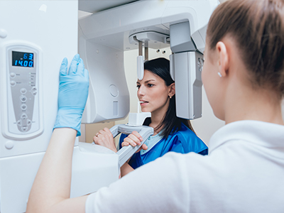
Digital radiography replaces traditional film with an electronic sensor that captures x-ray data and converts it into a digital image. The sensor—available in a variety of sizes and shapes—fits comfortably in the mouth or is positioned externally depending on the type of image needed. When the x-ray exposure occurs, the sensor records the pattern of x-ray photons and transfers that data to a connected computer for processing.

Software algorithms then convert the raw sensor data into a detailed, high-contrast image that can be adjusted for brightness, contrast, and magnification without needing a second exposure. Because the image is created digitally, the process eliminates chemical development and paper handling, producing an image that is both immediate and versatile. The result is a diagnostic-quality picture that helps clinicians identify cavities, bone loss, root issues, and other conditions with improved clarity.

Different sensors and imaging modes allow clinicians to capture bitewing, periapical, and panoramic-style images depending on the clinical need. Some systems also integrate with cone beam or intraoral camera data to give a more complete picture of oral structures. Together, these tools enable precise, evidence-based decision-making during routine exams and more complex restorative or surgical planning.

Speed and clinical collaboration: seeing results in real time

One of the defining advantages of digital radiography is immediacy. Images appear on a monitor almost instantly after exposure, allowing the dentist to review results in real time and discuss findings with the patient during the same appointment. This immediacy reduces uncertainty and helps patients understand their oral health through visual explanation, which often improves engagement and treatment acceptance.

Digital images can be annotated, enlarged, or adjusted to highlight areas of concern, making it easier for clinicians to explain diagnoses and proposed treatments. When multiple team members need to confer—whether hygienists, specialists, or lab technicians—the same image can be viewed simultaneously on different screens or shared digitally for consultation. That seamless collaboration shortens turnaround times and supports coordinated care plans.

For multidisciplinary cases, such as implant planning or complex restorative work, digital radiography integrates with planning software to allow precise measurements and guided workflows. This interoperability helps ensure that restorative components, surgical guides, and prosthetic designs align with the patient’s anatomy, reducing surprises and improving clinical predictability.

Patient safety: lower radiation exposure and optimized imaging

Digital radiography is designed to maximize the diagnostic value of each exposure while minimizing the dose of radiation a patient receives. Modern sensors are more sensitive than film, which means they require far less radiation to capture a usable image. Additionally, the ability to preview and adjust images digitally reduces the need for repeat exposures due to under- or overexposure.

Radiation safety remains a central concern in dental imaging, and digital systems are an important part of a multi-layered approach that includes proper shielding, collimation, and technique. Clinicians select the smallest field of view and lowest effective dose consistent with a clear diagnostic image, and imaging is performed only when clinically justified. These measures help keep cumulative exposure as low as reasonably achievable while still enabling accurate diagnosis.

For patients with heightened sensitivity concerns—such as children, pregnant patients, or individuals with complex medical histories—digital radiography offers flexible imaging protocols that balance diagnostic needs with conservative radiation practices. Clear communication about why an image is recommended and how exposure is minimized helps patients feel informed and reassured.

Recordkeeping, security, and seamless sharing of images

Because images are created and stored electronically, digital radiography simplifies recordkeeping and improves long-term accessibility. Every image can be linked directly to the patient’s chart, accompanied by notes, measurements, and timestamps, which supports efficient clinical documentation and follow-up care. Digital files are easier to archive and retrieve than physical film, helping practices maintain organized, complete records.

Secure image storage and transmission are vital. Contemporary dental offices use encrypted networks and secure practice management systems to protect patient information while enabling authorized sharing with specialists, referral partners, or outside labs when necessary. This controlled exchange speeds coordination while maintaining privacy and compliance with applicable regulations.

Having immediate access to historical images also benefits ongoing care: clinicians can compare current and past images side-by-side to monitor progression, evaluate treatment outcomes, or detect subtle changes over time. That continuity of information supports preventive strategies and helps clinicians intervene at the most appropriate moment.

Environmental advantages and practical benefits for office workflow

Switching from film-based radiography to digital systems eliminates the need for chemical developers, fixers, and paper—materials that require special handling and disposal. Removing these supplies reduces environmental impact, lowers hazardous waste generation, and streamlines daily operations. Many practices choose digital imaging in part because it aligns with broader sustainability goals.

From a workflow perspective, digital radiography reduces manual steps: there’s no film processing, no film storage boxes, and fewer instances of lost or damaged films. Staff can capture, label, and file images as part of a single digital task list, freeing time for patient care and reducing administrative bottlenecks. Faster image acquisition also shortens appointment times, improving patient comfort and practice efficiency.

Finally, the clarity and manipulability of digital images lead to more predictable clinical outcomes and fewer unnecessary follow-up visits. When imaging supports precise treatment planning, restorative and surgical interventions proceed with greater confidence, which benefits both patients and the practice’s operational rhythm.

In summary, digital radiography is a cornerstone of modern dental care—providing faster, safer, and more environmentally conscious imaging while supporting clearer communication and better clinical decisions. At Brian Howe DDS, Family Dentistry, we use these tools to enhance diagnostic accuracy and patient experience. If you’d like to learn more about how digital imaging informs your care, please contact us for additional information.

Frequently Asked Questions

What is digital radiography and how does it differ from traditional dental x-rays?

+

Digital radiography uses electronic sensors instead of film to capture x-ray data and create high-resolution images almost instantly. The sensor records the pattern of x-ray photons and transmits raw data to a computer where software converts it into a diagnostic image. At the office of Brian Howe DDS, Family Dentistry in Newark, OH, this approach replaces chemical processing and speeds clinical workflows.

Because images are digital, clinicians can adjust brightness, contrast, and magnification without additional exposures, improving diagnostic clarity. The elimination of film processing reduces handling steps and environmental waste while providing immediate visual feedback. This technological difference helps clinicians and patients review findings in real time during the same visit.

How are digital dental images captured and processed?

+

Images are captured by intraoral or extraoral electronic sensors that are placed in the mouth or positioned externally depending on the view required. When the x-ray exposure occurs, the sensor records the incoming photons and sends the data to connected imaging software for conversion and enhancement. Software algorithms then generate a high-contrast image that can be manipulated for optimal viewing.

Clinicians can annotate, measure, and enlarge regions of interest to support diagnosis and treatment planning without repeating exposures. Because processing is instantaneous, the team can confirm image quality immediately and retake only when clinically necessary. Integration with other digital tools, such as intraoral cameras or CBCT, further refines the diagnostic dataset.

What types of dental images can be obtained with digital radiography?

+

Digital radiography supports a range of image types including bitewing and periapical images for routine diagnostic needs as well as panoramic-style extraoral images for broader views. Many digital systems also integrate with cone beam computed tomography (CBCT) for three-dimensional imaging when more detailed anatomical information is required. This flexibility allows clinicians to select the appropriate view for each diagnostic question.

Different sensor sizes and imaging modes accommodate pediatric and adult patients as well as clinical scenarios such as endodontic assessment or periodontal evaluation. The ability to capture multiple image types within a unified digital environment streamlines recordkeeping and comparison. Clinicians use the specific image type that best answers the clinical question while minimizing exposure.

Is digital radiography safe and how is radiation exposure minimized?

+

Digital systems are more sensitive than traditional film, allowing clinicians to capture diagnostic images with substantially lower radiation doses. Modern practice follows the ALARA principle—keeping exposure as low as reasonably achievable—by selecting the smallest field of view and the lowest effective dose for each exam. Proper technique, shielding, and collimation are applied to further reduce patient exposure.

Because digital images can be previewed and adjusted, the need for repeat exposures is diminished, which lowers cumulative dose over time. Imaging is performed only when clinically justified, and practitioners tailor protocols for patients with special concerns. Clear communication about the purpose of imaging and the measures taken to limit exposure helps patients feel informed and reassured.

How does digital radiography improve diagnosis and treatment planning?

+

High-resolution digital images reveal subtle changes in tooth structure, bone levels, and root anatomy that aid earlier and more accurate diagnosis. Clinicians can magnify areas of interest, adjust contrast, and apply measurement tools to assess lesion size, bone loss, or root canal anatomy. These capabilities support evidence-based decision-making and reduce diagnostic uncertainty.

Digital files also facilitate side-by-side comparison with prior images, enabling clinicians to monitor progression or healing over time. When planning restorative or surgical treatment, digital imagery integrates with other digital records to create coordinated workflows. This precision leads to more predictable clinical outcomes and clearer patient communication about recommended care.

Can digital radiography be used for implant planning and complex restorative cases?

+

Yes, digital radiography is a key component of implant planning and complex restorative workflows, especially when integrated with three-dimensional imaging like CBCT and surgical planning software. Two-dimensional digital images provide initial assessment of bone levels and tooth positions, while CBCT offers volumetric data for evaluating bone volume, vital structures, and implant angulation. Together these tools support guided workflows and the design of surgical guides or prosthetic components.

Integration between digital images and planning software allows precise measurements and predictable placement of implants and restorative elements. This interoperability reduces the likelihood of intraoperative surprises and improves coordination among the restorative team, laboratory partners, and specialists. Careful preoperative imaging contributes to safer, more efficient procedures and better long-term outcomes.

How are digital images stored, shared, and protected to maintain patient privacy?

+

Digital images are linked directly to the patient record and stored in secure practice management or imaging systems that support encrypted storage and controlled access. Authorized staff can retrieve, annotate, and archive images while audit logs and user permissions help enforce privacy safeguards. When images must be shared with specialists or outside labs, secure transmission methods and encrypted file-sharing protocols are used to protect patient information.

Routine data management practices such as regular backups, secure network configurations, and software updates further protect clinical images from data loss or unauthorized access. Patients benefit from easy access to their imaging history and continuity of care when images are available for comparison. At Brian Howe DDS, Family Dentistry, these measures help ensure images are handled responsibly and in compliance with applicable privacy standards.

What should patients expect during a digital radiography appointment?

+

A typical digital radiography appointment is efficient and minimally invasive; an intraoral sensor or external imaging device is positioned briefly while a low-dose exposure is made. Patients may feel the sensor for a short time in the mouth, but modern sensors are available in sizes designed for comfort across age groups. The actual exposure is quick, and images appear on a monitor almost immediately for clinician review.

After images are captured, the dental team will explain what the images show and how they relate to the exam findings or recommended treatment. Because images can be enlarged and annotated, clinicians can guide patients through visual explanations that clarify diagnoses and options. This real-time review often reduces anxiety and supports informed decision-making.

Are there special considerations for children, pregnant patients, or medically complex patients?

+

Special populations require tailored imaging protocols that prioritize safety while meeting diagnostic needs, and clinicians adjust exposure settings, sensor choice, and field of view accordingly. For children, smaller sensors and reduced exposure parameters are used to limit dose while obtaining diagnostic images. For pregnant patients, imaging is performed only when essential and with appropriate shielding and justification.

Patients with complex medical histories may need interdisciplinary coordination and documentation to ensure imaging aligns with overall medical care. Clinicians discuss the risks and benefits of any recommended image and document the clinical rationale when special precautions are warranted. Open communication helps patients understand why imaging is advised and how exposure is minimized.

How does digital radiography benefit the environment and office workflow?

+

Digital radiography eliminates the need for chemical developers, fixers, and paper prints associated with film processing, reducing hazardous waste and the practice's environmental footprint. Removing these supplies simplifies supply management and avoids disposal requirements associated with chemical processing. Many practices choose digital imaging in part because it aligns with sustainability goals and reduces ongoing material consumption.

From a workflow standpoint, instant image capture and electronic filing shorten appointment times and reduce administrative steps like manual labeling and physical storage. Staff can capture, tag, and archive images as part of a single digital workflow, freeing time for patient care and improving operational efficiency. Patients typically benefit from quicker visits, clearer explanations, and more coordinated care when imaging supports precise treatment planning.

Contact Us

Our friendly and knowledgeable team is always ready to assist you. You can reach us by phone at (740) 344-4549 or by using the convenient contact form below. If you submit the form, a member of our staff will respond within 24–48 hours.

Please do not use this form for emergencies or for appointment-related matters.

SMS Consent
Submit

Thank You!

We appreciate you taking the time to visit our site. We'll review your message and be in touch with you soon.

Continue
The image shows a person s hands holding a smartphone while sitting at a table with a laptop open in front of them.