Existing Patients
(740) 344-4549
New Patients
(740) 212-1897

Intraoral cameras are compact, pen-sized imaging devices that capture high-resolution color images of teeth and soft tissues inside the mouth. Unlike traditional visual exams, these cameras reveal details that are difficult to see with the naked eye—hairline cracks, early decay between teeth, worn restorations, and subtle changes in the gum line. The clarity of the images helps clinicians detect issues earlier and with greater confidence, which supports more precise treatment decisions.
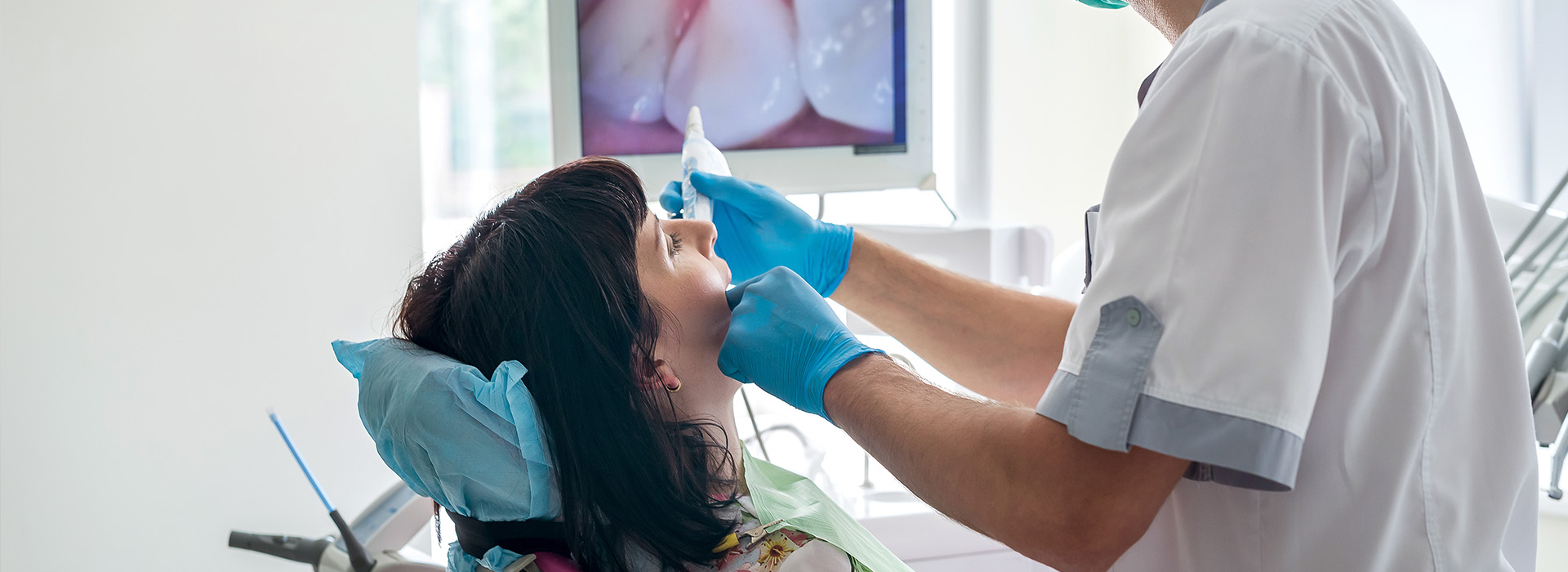
Beyond simply improving visualization, intraoral imaging transforms how information is shared during an appointment. Images appear in real time on a monitor, allowing patients to see exactly what the dental team sees. That shared visual reference creates a clearer foundation for discussion, so patients can make informed choices about their oral health without relying solely on verbal descriptions.
At Brian Howe DDS, Family Dentistry we incorporate intraoral photography into routine exams and targeted problem evaluations. The result is a diagnostic process that is both thorough and transparent: clinicians can document findings, explain treatment options using concrete visuals, and track changes over time through a reliable image record.
Using an intraoral camera is quick and comfortable. The device is gently guided into the mouth and positioned near the area of interest while the clinician captures a sequence of still images or a short video. Most patients experience little to no discomfort; the camera’s small size and smooth edges are designed for patient comfort and ease of access to hard-to-see areas.
Images captured with the camera are immediately displayed on a chairside monitor, giving patients a live view of their teeth and gums. This immediate visual feedback gives time to pause, ask questions, and discuss options while the condition is visible to everyone in the room. For many patients, seeing a clear image of an issue is more motivating than a verbal explanation alone.
Because the process is noninvasive and radiation-free, intraoral imaging can be used frequently as part of preventive care, progress checks, or pre-treatment evaluations. The quick capture time also minimizes chair time, letting clinicians gather the information they need without prolonging the visit unnecessarily.
Each image taken with an intraoral camera can be stored in a patient’s digital record and associated with clinical notes, radiographs, and treatment plans. These images create a visual timeline of oral health, making it easier to monitor the progression or resolution of conditions. When a clinician reviews a patient’s chart months or years later, these photographs provide valuable context that complements written observations and X-ray findings.
Stored intraoral images also improve collaboration with specialists and dental laboratories. Clear, annotated photographs can be shared with a periodontist, endodontist, or lab technician to illustrate specific concerns or to guide the fabrication of prosthetics. Because the images accurately depict anatomy and restorative needs, they reduce ambiguity and help ensure consistent communication across providers.
From an administrative perspective, maintaining a searchable image archive enhances record-keeping and clinical continuity. When follow-up care is needed, clinicians can quickly retrieve prior images to compare conditions over time, document treatment outcomes, and make data-driven recommendations for future care.
Intraoral cameras are designed with both safety and diagnostic performance in mind. They use visible light and do not emit ionizing radiation, which makes them safe for repeated use during exams and follow-ups. The camera housings are crafted for easy disinfection between patients, and many practices use disposable sleeves or strict sterilization routines to preserve infection control standards.
Advances in optics and sensor technology have significantly improved image fidelity. Modern intraoral cameras deliver high-resolution, true-color images with excellent depth-of-field, allowing clinicians to distinguish texture, color changes, and surface detail that matter clinically. Paired with chairside software, images can be enlarged, annotated, and compared side-by-side to show subtle changes that might otherwise go unnoticed.
The combination of high-quality imaging and secure digital storage also supports long-term care strategies. Clinicians can rely on reproducible images to measure wear, document lesion size, or monitor healing after treatment. Consistent imaging protocols produce dependable records, which in turn support accurate diagnoses and well-timed interventions.
Intraoral cameras are especially valuable in preventive dentistry because they make the early signs of disease visible and understandable. When patients can see discoloration, plaque accumulation, or the beginning of an enamel breakdown, they are often better equipped to take preventive steps—whether that means modifying home care, scheduling a focused hygiene visit, or consenting to a conservative restoration before the problem worsens.
During treatment planning, intraoral photos help clarify the rationale for different approaches. For example, clinicians can show the exact extent of a restoration failure or the margins of a crown that require attention, enabling a more precise conversation about options. This visual evidence can also help set realistic expectations for outcomes and timelines, leading to more collaborative decision-making.
Education is another significant benefit. Images captured during a routine exam can be used to highlight proper brushing and flossing targets, illustrate how restorations and natural teeth relate to one another, and explain the stages of periodontal health. When patient education is grounded in their own clinical images, the information often resonates more deeply and leads to better adherence to recommended care.
Ultimately, intraoral cameras are a tool for shared understanding: they empower clinicians to present accurate, compelling visual information and empower patients to participate actively in maintaining and improving their oral health.
In summary, intraoral cameras enhance diagnosis, communication, and long-term record-keeping while remaining safe and patient-friendly. They support preventive care and make treatment discussions more concrete and collaborative. If you’d like to learn more about how this technology is used in our office or how it could benefit your next visit, please contact us for more information.
An intraoral camera is a small, pen-sized imaging device that captures high-resolution color photographs and short videos of the teeth and soft tissues inside the mouth. The camera uses visible light and a tiny sensor to produce detailed images that reveal surface texture, color changes, cracks, and restoration margins that can be hard to see with the naked eye. Images are displayed in real time on a chairside monitor so both the clinician and patient can view the findings together.
During use the clinician guides the camera to the area of interest and captures stills or video segments that can be reviewed immediately. The process is noninvasive, radiation-free, and designed for quick capture to minimize chair time. Because the device produces true-color, magnified images, it supports clearer communication and more precise documentation of oral conditions.
Intraoral cameras enhance a standard visual exam by providing magnified, illuminated images that make small or subtle problems easier to detect. Hairline cracks, early interproximal decay, worn restorations, and minor gum-line changes often become visible on-camera long before they are obvious by sight or touch. These images help clinicians confirm findings and prioritize areas that need further investigation or intervention.
Because the images can be enlarged and compared side-by-side with prior photos, clinicians gain more confidence when recommending treatment or monitoring a condition. The visual evidence reduces ambiguity and supports objective decision-making rather than relying solely on verbal descriptions. This leads to earlier detection of problems and more targeted, minimally invasive care when appropriate.
Yes. Intraoral cameras are designed with patient comfort and safety in mind: they are small, smooth, and maneuverable so clinicians can access hard-to-see areas without causing discomfort. The devices use visible light and do not emit ionizing radiation, which makes them safe for repeated use during routine exams, progress checks, and pre-treatment evaluations. Many practices also use disposable sleeves or established disinfection protocols to maintain infection control between patients.
Most patients experience little to no discomfort during imaging, and the quick capture time helps keep visits efficient. The noninvasive nature of the technology makes it suitable for children, adults, and patients with sensitivity to traditional imaging methods. If a patient has specific concerns about comfort, the dental team can adapt technique and positioning to improve ease during the exam.
Images taken with an intraoral camera are stored in a patient’s digital chart and linked to clinical notes, radiographs, and treatment plans to create a comprehensive record. These photographs form a visual timeline that makes it easier to track progression or resolution of conditions over months and years. When clinicians review a chart later, the images provide concrete context that complements written observations and X-ray findings.
Stored images also support better follow-up care by making prior conditions easy to retrieve and compare to current findings. This searchable image archive improves record-keeping and continuity of care, allowing clinicians to document outcomes, monitor healing, and make data-driven recommendations during subsequent visits. Clear visual records also facilitate transparent discussions with patients about their oral health history.
Intraoral photos turn abstract descriptions into concrete visuals that patients can see and understand for themselves. When clinicians display a magnified image of a failing restoration, a crack, or an area of early decay, it creates a shared reference for discussing treatment options, expected outcomes, and timelines. This clarity helps patients make informed decisions based on what they can observe rather than relying only on verbal explanations.
Images can be annotated, enlarged, and compared with previous photos to illustrate why a particular approach is recommended and what improvements are realistic. That visual evidence also supports more precise treatment planning by showing exact margins, anatomy, and interrelationships between teeth and restorations. Overall, intraoral imaging fosters collaborative conversations and sets clearer expectations for care.
Absolutely. Because intraoral cameras reveal subtle surface changes and staining, they are a valuable tool for preventive dentistry and early detection of disease. Seeing plaque accumulation, early enamel breakdown, or the first signs of gum inflammation often motivates patients to adjust home care habits or schedule focused hygiene visits. Early identification of issues increases options for conservative treatment and can reduce the need for more invasive procedures later on.
Regular imaging during routine exams creates a visual baseline that makes it easier to notice small changes over time. Clinicians can use these comparisons to recommend targeted interventions, reinforce oral hygiene instruction, and monitor the effectiveness of preventive measures. By making early problems visible, intraoral cameras help patients take proactive steps to protect long-term oral health.
High-quality intraoral photographs provide a clear, shareable depiction of anatomy and restorative needs that enhances interdisciplinary communication. Clinicians can annotate and export images to a periodontist, endodontist, or laboratory technician to demonstrate specific concerns, margin details, or shade and contour relationships. These visuals reduce ambiguity and help specialists and technicians understand the clinical situation before the patient arrives or a restoration is fabricated.
Using consistent imaging protocols and attaching photos to referrals improves predictability and collaboration across providers. Clear images support accurate treatment sequencing, lab prescriptions, and pre-operative planning, which can streamline appointments and improve outcomes. When everyone involved sees the same visual information, coordination becomes more efficient and reliable.
The procedure is typically quick and straightforward: the clinician positions the small camera near the area of interest and captures a series of still images or a short video while you sit comfortably in the chair. Images appear immediately on a nearby monitor so you can review them with the clinician, ask questions, and see the exact concerns being discussed. The device’s compact size and smooth edges are intended to minimize discomfort and allow access to hard-to-see areas.
Because imaging is noninvasive and radiation-free, it can be repeated as needed during the same visit for documentation or comparison. The clinician may use the photos to annotate problem areas, explain treatment choices, or record baseline images for future monitoring. If you have any physical limitations or anxiety about the exam, let the team know and they will adapt their technique to make the process more comfortable.
Advances in optics, sensors, and chairside software have significantly increased image fidelity, color accuracy, and depth of field in modern intraoral cameras. Contemporary devices capture high-resolution, true-color images that reveal fine surface detail and subtle color variations important for diagnosis. Paired with software, clinicians can enlarge, annotate, compare, and securely store images, turning single photos into useful diagnostic tools and reliable records.
Improved ergonomics, smaller form factors, and better illumination have also made cameras easier to use on a wide range of patients and in challenging intraoral positions. Enhanced connectivity and secure digital storage streamline integration with electronic health records, enabling efficient retrieval and sharing when coordinating care. These technological improvements increase the utility of intraoral imaging for both diagnosis and long-term patient management.
At the office of Brian Howe DDS, Family Dentistry, intraoral cameras are incorporated into routine exams and problem-focused visits to improve visualization and patient understanding. Clinicians document findings with clear photographs, review those images with patients on a chairside monitor, and use the visuals to explain treatment options and monitor changes over time. This approach supports transparent communication and helps patients participate actively in care decisions.
Stored images become part of the digital record and are used to track healing, compare outcomes, and coordinate care when referrals or laboratory work are needed. In our Newark, Ohio office, the team follows standard infection control and imaging protocols to ensure images are reliable, reproducible, and useful for long-term oral health planning. If you would like more details about how imaging is used during your visit, the team can walk you through the process during your next exam.
Our friendly and knowledgeable team is always ready to assist you. You can reach us by phone at (740) 344-4549 or by using the convenient contact form below. If you submit the form, a member of our staff will respond within 24–48 hours.
Please do not use this form for emergencies or for appointment-related matters.
